

Guides
3D Image Acquisition using Structured-Light Scanners
Updated on July 25, 2024
Craniofacial Identification techniques, such as craniofacial superimposition (CFS), craniofacial reconstruction (CFR) and others are based on the use of 2D or 3D images of the face and skull [1] to try to establish the identity of an individual. Historically, these procedures have been carried out only with photographs due to the lack of the necessary tools for the acquisition of three-dimensional images [2],however, in recent decades, advances and new developments in this field have made it possible both to obtain high-quality 3D models and to make this technology accessible to the general public. Because of this, it is currently recommended that the techniques aforementioned are carried out using 3D post-mortem models [3], and not 2D post-mortem photographs, as these offer a series of advantages when performing the AM-PM comparison and superimposition processes –- making it possible to align, estimate the pose, scale and adjust the perspective of the model to improve its coincidence with the AM image –.
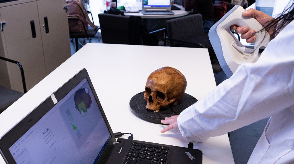
For the acquisition of three-dimensional images in anthropology, whether of skulls or faces, we can use different devices – whose digitization technologies can be very diverse [4] – depending on the task performed by the professional in question. Of these, structured light scanners are of particular interest to our field, as they offer several advantages over other available options, including portability and compact size – which gives us not only greater flexibility of movement when scanning, but also the possibility of working on battery power and therefore on the ground –; the versatility they offer, so that they can be used both on tripods and manually with free movement, thus increasing the range of scannable sample sizes, as the field of view is not limited to a fixed position [5] and, finally, their scanning speed, as this is considerably reduced when compared to digitisations performed using other techniques [6].
The scanning process with these devices is intuitive and does not require more than basic initial training, which will help us to better understand the scanner, the associated software and its possibilities. This training will vary slightly depending on the brand or manufacturer of our equipment, however, in general, we will achieve a good result as long as we follow a series of specific steps during the scanning process:
- In the first place, it is necessary to check that the scanner is correctly connected and configured with a computer, in which the corresponding software must also be installed. After this, we must perform a calibration according to the workplace, adjusting values such as scanning field, focal length and resolution, following the manufacturer’s instructions.
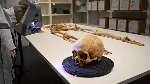
- We will set up the work area in a well-lit place and make sure that there are no shadows that could affect the quality of the process [7], then we will place the object to be scanned on a rotating base or on a smooth surface depending on the morphology and size of the object. Rotating bases – whether automated or manual – are very useful because, by rotating the base and not the person operating the scanner, they allow the geometry of the object to be captured in a more fluid way, without the worry of losing track of the piece. It may be necessary to combine several scans for the same sample – with and without a rotating base, in different positions, and even with free movement or with a tripod – in order to acquire more detailed information of the most problematic regions, such as the orbital region, the piriform aperture and the zygomatic arches, just to mention a few examples that we would find in a skull.
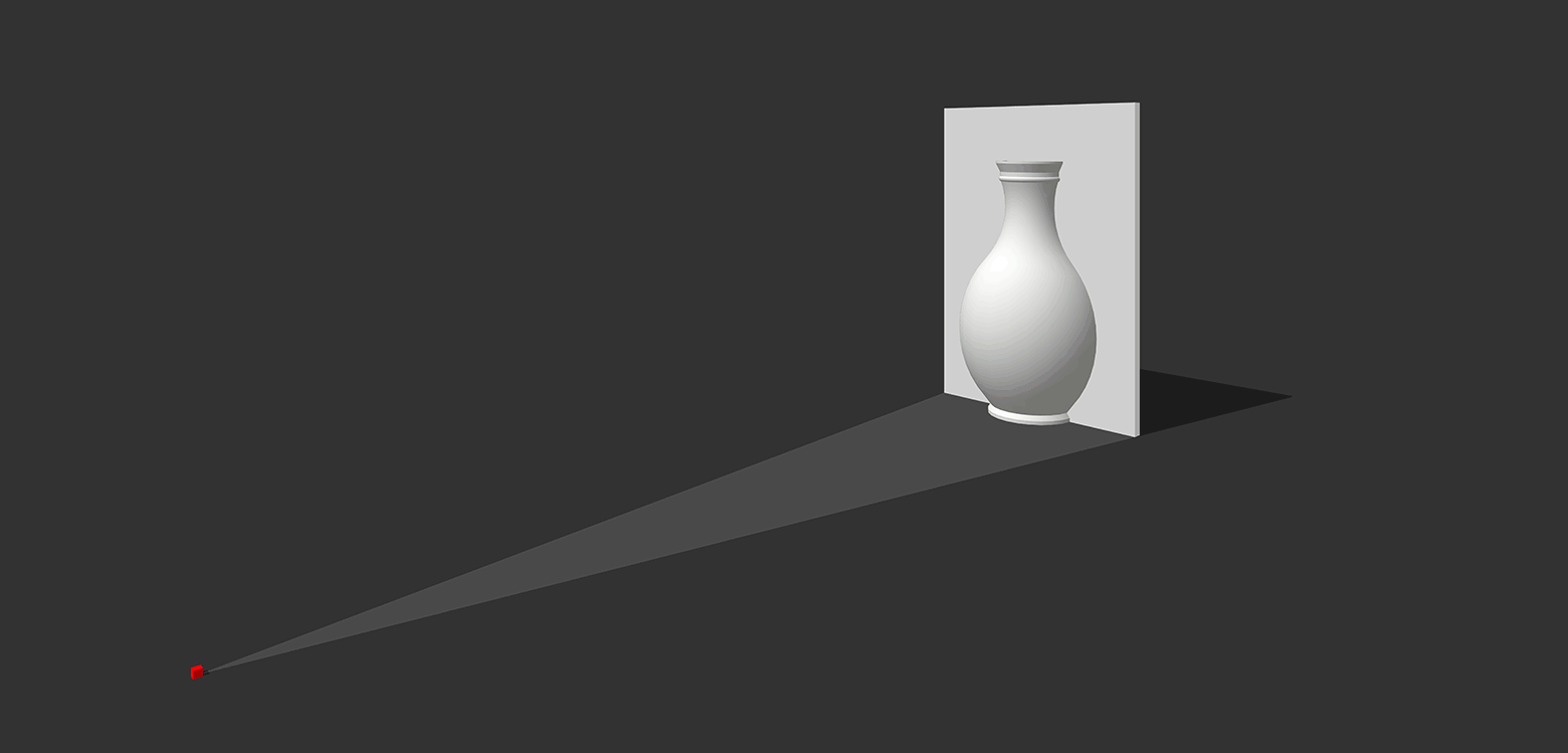
- The scanner will start projecting a structured light pattern – usually composed of linear or circular motifs, whose geometry is perfectly known – onto the object after making sure that the calibration has been performed. The scanner’s camera will take images of the illuminated object with the projected light patterns, which will contain information about the deformation of its design caused by the shape of the object [8]. Beyond capturing the geometry of the object, there are scanners on the market that record its texture – a bitmap linking the color of each pixel to its corresponding point on the surface of the 3D model – by having an additional camera.
- The scanner software will process the data after capturing the images and use sophisticated algorithms to analyze the light deformation patterns and reconstruct the shape of the object in three dimensions, giving us a complete and detailed visual representation of the model.
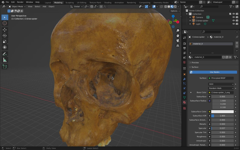
- The model obtained will need post-processing to remove unwanted artifacts – the base on which the model was scanned, objects that interfered with the scene, etc. –, merge the generated meshes in case we have made more than one scan for the same sample and simplify the polygons of the final model without losing the details. The tools for this processing are usually included in the scanner software itself, although there are also open-source alternatives, such as MeshLab, Blender and others. Once the model has been processed, it will be exported in STL, OBJ or PLY format. The last two are the most recommended for anthropological work because they contain both the geometry of the object and its texture.
During the scanning process, if the calibration or environmental conditions were not correct, we may encounter a series of errors such as the appearance of “noise” or distortions in the mesh; the lack of detail in areas that are difficult for the scanner to reach, such as the inside of the orbits in the case of a skull; the oversimplification of the geometry of the object – causing, for example, that the cranial sutures can’t be properly appreciated – or the excessive size of the files generated, among others.
Given the advantages offered by 3D scanners, both for its reduced learning curve and for other characteristics, including its size and portability, as well as its price, we recommend that, when choosing a device and particularly for Craniofacial Identification, it should have an accuracy – deviation allowed for each point of the captured cloud with respect to the real position of the object – of between 0.05 and 0.1 mm; a resolution – minimum distance between the scanned points, so that the higher the resolution, the greater the density of the cloud – of between 0.1 and 0.25 mm and a scanning speed – speed with which the scanner captures the points – of between 5 and 10 fps. Standards such as MEPROCS [9] placeparticular emphasis on the need for scanner accuracy to never exceed 1mm, and for equipment to be fitted with a texture capture device.
At Panacea, we are used to working with structured light scanners and we keep up to date with the latest advances, developments and innovations in digitization and generation of three-dimensional models. Below there’s a list of publications that you can consult to learn more about the subject.
If you have other questions about the use of this equipment in anthropology and forensic sciences, do not hesitate to contact us and we will be happy to give you a helping hand.

Verónica Martínez
Verónica is bioanthropologist and researcher at Panacea. She studied Archaeology at the University of Granada and later completed her Master’s degree in Physical and Forensic Anthropology.
In recent years she has been involved in projects as an osteoarchaeologist and anthropologist, working on the excavation and study of mass graves from the Spanish Civil War.

Guillermo Ramírez
Guillermo is a physical and forensic anthropologist and researcher at Panacea Cooperative Research. He holds a degree in biology from the University of Granada, having later complemented his studies with a Master’s degree in Physical and Forensic Anthropology at the same university and another Master’s degree in Anthropology and Forensic Genetics from the Fundación General Universidad de Granada.
[1] C. Stephan, ‘Craniofacial identification: techniques of facial approximation and craniofacial superimposition’, 2009, pp. 304–321.
[2] M. Yoshino, ‘Craniofacial superimposition’, in Craniofacial Identification, C. Wilkinson and C. Rynn, Eds., Cambridge University Press, 2012, pp. 238–253. doi: 10.1017/CBO9781139049566.019.
[3] O. Ibáñez et al., ‘MEPROCS framework for Craniofacial Superimposition: Validation study’, Leg. Med., vol. 23, pp. 99–108, 2016.
[4] M. T. Ross, R. Cruz, T. L. Brooks-Richards, L. M. Hafner, S. K. Powell, and M. A. Woodruff, ‘Comparison of three-dimensional surface scanning techniques for capturing the external ear’, Virtual Phys. Prototyp., vol. 13, no. 4, pp. 255–265, 2018.
[5] R. M. Ruiz, M. T. M. Torres, and P. S. Allegue, ‘Comparative analysis between the main 3d scanning techniques: Photogrammetry, terrestrial laser scanner, and structured light scanner in religious imagery: The case of the holy christ of the blood’, ACM J. Comput. Cult. Herit. JOCCH, vol. 15, no. 1, pp. 1–23, 2021.
[6] J. Shan, Z. Li, D. Lercel, K. Tissue, J. Hupy, and J. Carpenter, ‘Democratizing photogrammetry: an accuracy perspective’, Geo-Spat. Inf. Sci., pp. 1–14, 2023.
[7] P. Rachakonda, B. Muralikrishnan, and D. Sawyer, ‘Sources of errors in structured light 3D scanners’, in Dimensional Optical Metrology and Inspection for Practical Applications VIII, SPIE, 2019, pp. 25–37.
[8] J. Geng, ‘Structured-light 3D surface imaging: a tutorial’, Adv. Opt. Photonics, vol. 3, no. 2, pp. 128–160, 2011.
[9] S. Damas, O. Cordón, and O. Ibáñez, ‘MEPROCS Craniofacial Superimposition Framework’, in Handbook on Craniofacial Superimposition: The MEPROCS Project, Cham: Springer International Publishing, 2020, pp. 139–152. doi: 10.1007/978-3-319-11137-7_8.
[10] ‘How does structured-light 3D scanning work? | Professional 3D scanning solutions | Artec 3D’. https://www.artec3d.com/learning-center/structured-light-3d-scanning (accessed May. 28, 2023).
[11] M. I. Huete, O. Ibáñez, C. Wilkinson, and T. Kahana, ‘Past, present, and future of craniofacial superimposition: Literature and international surveys’, Leg. Med., vol. 17, no. 4, pp. 267–278, 2015, doi: https://doi.org/10.1016/j.legalmed.2015.02.001.
[12] A. Norouzi et al., ‘Medical image segmentation methods, algorithms, and applications’, IETE Tech. Rev., vol. 31, no. 3, pp. 199–213, 2014.
[13] R. C. Nightingale, M. T. Ross, M. C. Allenby, M. A. Woodruff, and S. K. Powell, ‘A method for economical smartphone-based clinical 3D facial scanning’, J. Prosthodont., vol. 29, no. 9, pp. 818–825, 2020.
[14] E. Rakitina, I. Rakitin, V. Staleva, F. Arnaoutoglou, A. Koutsoudis, and G. Pavlidis, ‘An overview of 3D laser scanning technology’, in Proc. of the International Scientific Conference,(Varna, Bulgaria), 2008.