

Introductions
A quick introduction to Biological Profile Estimation
Biological Profile
Published on October 24, 2022
Written by Stefano De Luca
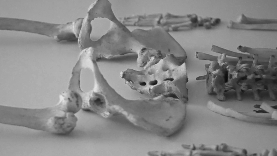
Forensic anthropology is the application of biological anthropology and its methods to identify the living and the dead, especially in the framework of a legal investigation of human remains (1). Reconstructing the biological profile may help law enforcement, medical examiners, and medicolegal death investigators narrow down the list of potentially unidentified persons by automatically excluding a large percentage of the population (2,3).
Estimating the biological profile is an attempt to reconstruct an individual’s life and death, including biological sex and age, ethnicity, stature, and any individualizing skeletal or dental traits. In the case of subadult skeletal remains (fetal, infant, child, and other immature individuals under the age of 18), it is noteworthy that the only parameter of the biological profile that is routinely estimated is age (3).
Sex Estimation
Biological sex is one of the first parameters analyzed when estimating the biological profile. Other steps, such as ethnic origin and stature estimations, rely on an accurate and precise assessment of sex (2,3). It is essential to understand that it is based on the concept of sexual dimorphism, that is, the existence of appreciable differences between males and females of a certain species, within and between populations, according to size and shape (3).
Current methods used to estimate sex from human remains consist of either morphological or qualitative (form or structure) and metric or quantitative (measured) traits analyses (7).
Concerning the morphological differences, the assumption is that the more gracile expression should have a greater frequency in females, while the more robust expressions should be more common in males (8). So, a particular feature or a skeletal region needs to be visually examined to verify if it is robust/gracile or, in some cases, present/absent. In this case, the skull and the pelvis are the most commonly used areas of the skeleton for estimation (8). These differences are related to sexual dimorphism usually varying in the amount of robusticity observed between males and females (2,3,8). Regarding the metric traits, this approach, which can include multiple (multivariate) or single (univariate) measurements from several skeletal elements, focuses on sexually dimorphic size (breadth and length of different bones and teeth) differences between the sexes (2,3,9).
Age Estimation
In the human identification process, estimating biological age may be used to determine whether an individual should be included or excluded from further legal consideration on the basis of the consistency between the known age and estimated age (2,3).
In forensic cases, the application of the most accurate methods depends on what skeletal elements are preserved and what general age is represented (3). In general, techniques applied to estimate age in subadults (i.e., not yet adult) differ from those required to analyse the skeleton of an adult individual, being then relied on the evaluation of two fundamental physiological processes: 1) growth and development in subadults, and 2) degeneration (or aging) in adults.
During growth and development, estimating age is more accurate and reliable as the biological–chronological relationship is clearly reflected in the subadult skeleton, which grows according to predictable rates (3). In this case, two methods are commonly used: 1) the analysis of the epiphyseal union, and 2) the assessment of dental development and maturation (2,3).
After our skeleton ceases to grow, the bone, joint surfaces, and teeth begin to undergo structural changes, or degeneration, associated with aging. This process begins normally after 18 years of age, with more robust changes after middle adulthood (after 35 years of age). This does not happen at such predictable rates and, therefore, results in less accurate and precise intervals of estimated age (2,3).
Regarding the most used methods, different skeletal and dental regions may be examined to estimate age in adults and numerous aging methods for the same skeletal region/s are available (3). The pubic symphysis is one of the most reliable areas of the skeleton for adult age estimation (3,11). Other methods include changes in the morphology of the auricular surface (pelvis) (3,11) and the sternal rib end (3,11). As regards the dental age, the most frequently considered parameters are dentine translucency (10,12) and secondary dentine deposition (10,13). Although many of these methods have been already tested and frequently used in forensic cases, there is no standardized way of combining multiple age estimation methods into a final age estimate to report to public prosecutors, officials, and courts of law (2,3).
Stature Estimation
Stature estimation is of utmost importance for the identification of skeletonized and decomposed dead bodies (2,3). Nearly every individual has their biological height recorded on personal documents. However, forensic anthropologists calculate an interval that is only an approximation of an individual’s true height (2,3).
The most used method is the regression model which requires measuring a limb bone length according to the sex, age, and ethnicity of the analyzed subject to examine the relationship between height and bone length in order to calculate a prediction interval to estimate stature (2,3). This process is performed on long bones such as the humerus, femur, and tibia. If these bones are unavailable, the ulna, radius, and fibula can also provide a good interval for the expected stature (3). This interval is normally expressed as a set of two numbers: the first one is the mean stature while the second one is a margin of error, which provides the upper and lower limits of the mean (e.g., 180 ± 5 cm). To accommodate the interval of variation, 90% or 95% confidence intervals are generally applied (14). For most standards, the limb bone measurements are usually maximum lengths. In addition to formulae for individual limb bones, there are also regression formulae for multiple bones (15).
Eventually, fragmented limb bones and some non-limb bones (e.g., skulls, pelvis, and bones of the hands or feet) may be used in regression formulae to estimate stature (15).
Ethnicity Estimation
Ethnicity, like the other parameters of the biological profile, is estimated to facilitate the identification of an unknown individual by helping to narrow the pool of possible matches with missing persons (2,3).
Nowadays, ethnicity is often incorrectly labeled as race (16). However, this concept is not valid on a biological level, and forensic anthropologists have attempted to reframe this parameter of the biological profile. Instead of using race, the words ancestry or ethnicity, as an expression of our phenotype (outward appearance), are employed in casework (16,17). Because our phenotype may vary according to environmental action, it can be estimated by analyzing the morphological traits of the skeletal remains (16,17).
As already stated, when a person is reported as missing, the information gathered by law enforcement includes age, biological sex, stature, and ethnic origin. So, since human identification is a comparative process, all these parameters should be used in forensic case analysis in order to obtain at least an exclusion. In addition, some authors (17) suggest that ethnicity should be the first parameter to be estimated because the results will determine which method should be used when the other three identification parameters are evaluated.
In general, anthropologists are able to divide humans into broad geographically discrete groups, including (but not limited to) the following: European, African, Asian, Native American, and Hispanic (18).
Traditionally, ethnic origin estimation was achieved by a visual examination (macromorphoscopic or MMS study) of some elements of the human skull (e.g., interorbital breadth, nasal aperture width, or the morphology of the nasal spine, palatine sutures). However, in order to reduce subjectivity, the majority of the methods focus now on a list of nonmetric cranial MMS traits precisely determined within a statistical framework to allow the practitioners to generate a statistically validated prediction of geographic origin (19). The initial isolation and standardization of cranial MMS traits are provided by Hefner (20-22), and Hefner and Linde (23). Reported rates of inter- and intra-observer agreements are available.
Written by
Dr. Stefano De Luca
Stefano De Luca is a Marie Sklodowska-Curie experienced researcher at the Panacea Cooperative Research S. Coop., where he leds the project “UMAFAE – Unaccompanied Minors Automatic Forensic Age Estimation”, funded by the European Union. He holds an archaeology degree from the University of Salento (Lecce, Italy) and an MSc degree in Physical Anthropology, and a Ph.D. degree in Biomedicine from the University of Granada (Spain). He was a forensic expert at the Unidad de Derechos Humanos (Servicio Médico Legal, Santiago de Chile); he is also a visiting professor at the Universidad de la Frontera (UFRO, Temuco, Chile).

Stefano de Luca is a member of the AgEstimation Project, led by the University of Molise (Campobasso, Italy), which aimed to develop quantitative methods to estimate age in immature and adult subjects. He is the author of more than 50 scientific papers, published in several high-impacted international journals in the fields of forensic sciences and legal medicine. He is the author and co-author of numerous book chapters and he is also a reviewer and editor of some scientific international journals of the forensic and legal fields. The main focus of his research is the development and validation of dental and skeletal age estimation, and the automatization of the main dental and skeletal techniques to assess the legal age of undocumented, unaccompanied, and separated minors.
References
1. Randolph-Quinney, P.S., Mallett, X., Black, S.M. (2009). Forensic Anthropology. In A. Jamieson, A. Moenssens (eds.) Wiley Encyclopedia of Forensic Science. London: John Wiley and Son Ltd. Pp 1-27.
2. Márquez-Grant, N., Roberts, J. (2021) Redefining forensic anthropology in the 21st century and its role in mass fatality investigations. European Journal of Anatomy, 25(2):19-34.
3. Hackman, L., Delabarde, T., Roberts, J., de Boerr, H. (2022). Forensic Anthropology: a primer for courts. The Royal Society.
4. Champod, C., Chamberlain, P. (2009). Fingerprints. In J. Frasier, R. Williams (eds.) Handbook of Forensic Science. London: Routledge. Pp 57-83.
5. Dumache, R., Ciocan, V., Muresan, C., Enache, A. (2016). Molecular DNA Analysis in Forensic Identification. Clinical Laboratory, 62(1-2):245-248.
6. David, T.J., Lewis, J.M. (eds.) (2018). Forensic Odontology. Principles and Practice. London: Academic Press, Elsevier, pp. 338.
7. Christensen, A.M., Passalacqua, N.V., Bartelink, E.G. (2014). Forensic Anthropology: Current Methods and Practice. San Diego, CA: Elsevier.
8. Spradley, M.K., Jantz, R.L. (2011). Sex estimation in forensic anthropology: skull versus
postcranial elements. Journal of Forensic Sciences, 56:289–296.
9. Albanese, J., Eklics, G., Tuck, A. (2008). A metric method for sex determination using the proximal femur and fragmentary hipbone. Journal of Forensic Sciences. 53:1283–1288.
10. Stavrianos, C., Mastagas, D., Stavrianou, I., Karaiskou, O. (2008). Dental age estimation of adults: a review of methods and principals. Research Journal of Medical Sciences. 2(5):258–268.
11. Garvin, H.M., Passalacqua, N.V., Uhl, N.M., Gipson, D.R., Overbury, R.S., Cabo, L. 2012). Developments in Forensic Anthropology: Age-at-Death Estimation. In D.C., Dirkmaat (ed.). A companion to forensic anthropology. First edition. West Sussex: Wiley-Blackwell. Pp. 202-223.
12. Prince, D.A., Ubelaker, D.H. (2002). Application of Lamendin’s adult dental aging technique to a diverse skeletal sample. Journal of Forensic Sciences. 47(1):107–116.
13. Cameriere, R., De Luca, S., Alemán, I., Ferrante, L., Cingolani, M. (2012). Age estimation by pulp/toot ratio in lower premolars by orthopantomography. Forensic Science International. 214(1-3):105-112.
14. De Mendonça, M.C. (2000). Estimation of height from the length of long bones in Portuguese adult population. American Journal of Physical Anthropology. 112(1):39-48.
15. Willey, P. (2014). Stature Estimation. In C., Smith (ed.) Encyclopedia of Global Archaeology. New York: Springer.
16. Albanese, J., Saunders, S.R. (2006). Is it possible to escape racial typology in forensic identification? In A., Schmitt, E., Cunha, J., Pinheiro (eds.). Forensic Anthropology and Medicine. Totowa, NJ: Humana Press. Pp. 281–316.
17. Algee-Hewitt, B.F.B., Coelho, C., Navega, D., Cunha, E. (2020). Statistical approaches to ancestry estimation: new and established methods for the quantification of cranial variation for forensic casework. In Z., Obertova, A., Stewart, C., Cattaneo (eds.). Statistics and probability in Forensic Anthropology. London: Academic Press.
18. Algee-Hewitt, B.F.B. (2016). Population inference from contemporary American craniometrics. American Journal of Physical Anthropology. 160:604–624.
19. Hefner, J.T., Ousley, S.D. (2014). Statistical classification methods for estimating ancestry using morphoscopic traits. Journal of Forensic Sciences. 59:883–890.
20. Hefner JT. The utility of nonmetric cranial traits in ancestry determination – Part II. Paper presented at the 55th Annual Meeting of the American Academy of Forensic Sciences, Chicago, IL, 2003.
21. J., Hefner. (2007). The statistical determination of ancestry using nonmetric traits. Dissertation, University of Florida.
22. Hefner, J. (2009). Cranial nonmetric variation and estimating ancestry. Journal of Forensic Sciences. 54(5):985-995.23. Hefner, J., Linde, K.C. (2018). Atlas of human cranial macromorphoscopic traits. London: Academic Press.