

Introductions
A quick introduction to Comparative Radiography
Comparative Radiography
Published on July 4, 2022
Written by Pablo Mesejo
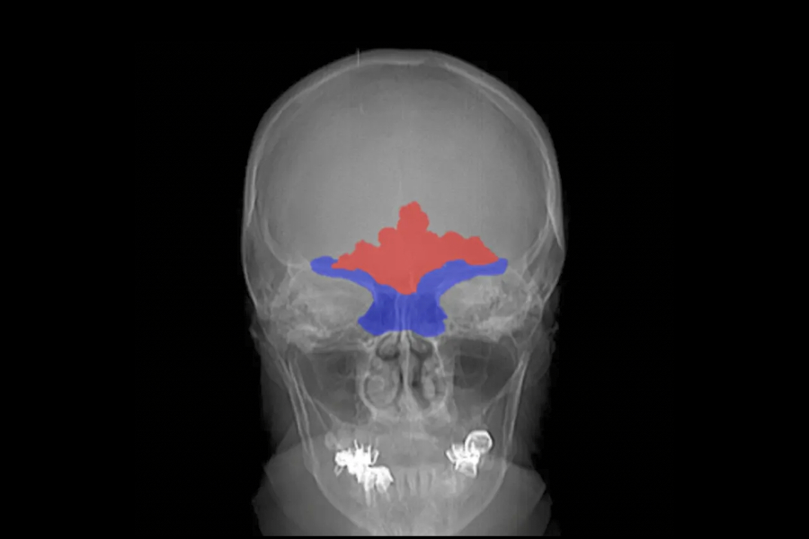
Comparative radiography (CR) [1] is a forensic identification technique based on the comparison of skeletal structures in ante-mortem (AM) and postmortem (PM) radiographic images. In the forensic literature, several skeletal structures have been reported as useful for candidate shortlisting or positive identification using CR, based on their individuality and uniqueness [2]. The most commonly employed skeletal structures in CR are located in the skull, chest, and thoracic areas. In the skull, the most frequently used are teeth [3], frontal sinuses [4], and the cranial vault [5]. In the chest and thoracic areas, clavicles [6] and vertebral features [7] are those most commonly considered. There are also a few bones outside these areas that are traditionally used, such as the bones of the hand [8] and the patella [9].
The advance in medical imaging techniques naturally led to the comparison of skeletal structures using all kinds of medical images. Depending on the dimensionality of the images compared, we can distinguish three different paradigms:
- 2D AM Radiograph – 2D PM Radiograph: This comparison methodology [18,19] consists in manually generating PM radiographs of the deceased, reproducing the acquisition protocol, and the pose of the targeted skeletal structure of the candidate AM radiographs. Once the PM radiographs have been acquired, AM and PM radiographs are visually compared looking for consistences, explainable inconsistencies (e.g. resulting from the time elapsed among AM and PM radiographs, degenerative processes, the effect of gravity on the body, perspective distortions resulting from errors reproducing the AM radiographs, etc.) and inconsistent findings (e.g. point-to-point comparison of frontal sinuses using a standardized protocol [18]). This 2D-2D comparison is the most common and extended approach.
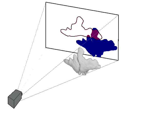
- 2D AM radiograph – 3D PM images: requires the generation of PM simulated radiographs from a 3D image (previously acquired using a 3D scan: CT, CBCT, MRI, laser or light structure, etc.) to simulate the AM radiographs [19,20,21,22]. These simulated radiographs are normally obtained by the forensic anthropologist through a trial-and-error process using generic medical imaging software. Alternatively, recent AI-based approaches automate this trial-and-error process [9, 32-38] (see figure). In any case, AM radiographs and PM simulated radiographs are visually compared as in the radiograph-radiograph approach. Additionally, a matching score is provided by automatic methods based on the assessment of bone morphology.
- 3D AM Image – 3D PM Image: The comparison is performed via visual comparison of their 3D shapes [17], avoiding occlusions or perspective distortions, or via anthropological measurements [24], where the distances can be directly compared since 3D models maintain the original physical units. This comparison methodology [25,26,27,28] is the most reliable one since 3D images retain more information about the skeletal structure than 2Dimages. Furthermore, this methodology avoids errors related to the acquisition of the PM radiographs.
In practice, the most common identification scenarios are based on the comparison of AM radiographs against PM radiographs or 3D images, since the availability of AM radiographs is more frequent [29]. However, the acquisition process of a PM radiograph “simulating” the AM one is a complex and error-prone trial-and-error process since small changes in the acquisition parameters result in great changes in the skeletal structures’ silhouettes [31]. Furthermore, both the acquisition of the PM radiographs and their visual comparison against the AM ones rely entirely on the forensic expert’s skills and experience. Consequently, the utility of these methods is reduced because of the time required (2-8 hours per superimposition depending on the AM radiograph [15]), the inherent subjectivity, and the errors related to the analyst’s fatigue.
The use of the 3D image methodology has become more popular in recent years since an increasing number of forensic labs have access to CT scans (via agreements with hospitals or acquiring a dedicated CT) and/or 3D surface scanners [30], thanks to their great availability and relatively low cost. Within the 2D-3D scenario, automatic comparative radiography methods overcome all the drawbacks 2D-2D approaches have corning time, accuracy, and subjectivity. The case of 3D-3D, although being the most reliable, becomes the most difficult to be applied as it requires a 3D AM image, which is still not as common as a 2D radiograph.
Written by:
Dr. Pablo Mesejo
Dr. Pablo Mesejo is a founding partner of Panacea and a permanent professor at the University of Granada. Previously, he was a postdoctoral researcher (2020-21) and Marie Curie Experienced Researcher (2018-2020) at the same institution, where he led the EU project “Soft Computing and Computer Vision for Comparative Radiography in Forensic Identification”.

He also held positions at Inria Starting Research (France, 2016-18) and postdoctoral researcher at Inria (France, 2014-16), a postdoctoral researcher in a laboratory belonging to the CNRS (France, 2013-14), and Marie Curie predoctoral researcher at the University of Parma (Italy, 2010-13).Dr. Mesejo’s scientific career has produced 30 publications in indexed journals and top-level international conferences. The main focus of his research is the analysis and design of machine learning, computer vision, and computational intelligence methods mainly to solve biomedical image analysis problems.
Publications
1. M. J. Thali, B. Brogdon, M. D. Viner. Forensic Radiology, CRC Press, 2002.
2. T. Kahana, J. Hiss. Identification of human remains: forensic radiology, Journal of Clinical Forensic Medicine 4 (1) (1997) 7-15.
3. A. Pretty. A look at forensic dentistry-part 1: The role of teeth in the determination of human identity, British Dental Journal 190 (7) (2001) 359-366.
4. G. Quatrehomme, P. Fronty, M. Sapanet, G. Grevin, P. Bailet, A. Ollier. Identification by frontal sinus pattern in forensic anthropology, Forensic Science International 83 (2) (1996) 147-153.
5. A. B. Maxwell, A. H. Ross. A radiographic study on the utility of cranial vault outlines for positive identifications, Journal of Forensic Sciences 59 (2) (2014) 314{318.
8. M. G. Koot, N. J. Sauer, T. W. Fenton. Radiographic human identification using bones of the hand: A validation study, Journal of Forensic Sciences 50 (2) (2005) 263-268.
9. E. Niespodziewanski, C. N. Stephan, P. Guyomarc’h, T. W. Fenton. Human identification via lateral patella radiographs: A validation study, Journal of Forensic Sciences 61 (1) (2016) 134-140.
15. E. Streetman, T. W. Fenton. Comparative medical radiography: Practice and validation, in: K. E. Latham, E. J. Bartelink, M. Finnegan (Eds.), New Perspectives in Forensic Human Skeletal Identification, Academic Press, 2018, pp. 251-264.
16. T. D. Ruder, C. Brun, A. M. Christensen, M. J. Thali, D. Gascho, W. Schweitzer, G. M. Hatch. Comparative radiologic identification with CT images of paranasal sinuses – development of a standardized approach, Journal of Forensic Radiology and Imaging 7 (2016) 1-9.
17. D. Gibelli, M. Cellina, A. Cappella, S. Gibelli, M. M. Panzeri, A. G. Oliva, G. Termine, D. De Angelis, C. Cattaneo, C. Sforza. An innovative 3D-3D superimposition for assessing anatomical uniqueness of frontal sinuses through segmentation on CT scans, International Journal of Legal Medicine 133 (4) (2019) 1159-1165.
18. A. H. Ross, A. K. Lanfear, A. B. Maxwell. Establishing standards for side-by-side radiographic comparisons, The American Journal of Forensic Medicine and Pathology 37 (2) (2016) 86-94.
19. T. Kahana. El aporte de la radióloga al avance de la antropología forense: perspectiva profesional (in spanish), Ph.D. thesis, University of Granada (2009).
20. G. M. Hatch, F. Dedouit, A. M. Christensen, M. J. Thali, T. D. Ruder. RADid: A pictorial review of radiologic identification using postmortem CT, Journal of Forensic Radiology and Imaging 2 (2) (2014) 52-59.
21. M. Pfaei, P. Vock, R. Dirnhofer, M. Braun, S. A. Bolliger, M. J. Thali. Post-mortem radiological CT identification based on classical ante-mortem x-ray examinations, Forensic Science International 171 (2-3) (2007) 111-117.
22. N. Shinkawa, T. Hirai, R. Nishii, N. Yukawa. Usefulness of 2D fusion of postmortem CT and antemortem chest radiography studies for human identification, Japanese Journal of Radiology 35 (6) (2017) 303-309.
24. D.-I. Kim, U.-Y. Lee, S.-O. Park, D.-S. Kwak, S.-H. Han. Identification using frontal sinus by three-dimensional reconstruction from computed tomography, Journal of Forensic Sciences 58 (1) (2013) 5-12.
25. M. Iino, H. Fujimoto, M. Yoshida, H. Matsumoto, M. Q. Fujita. Identification of a jawless skull by superimposing post-mortem and ante-mortem ct, Journal of Forensic Radiology and Imaging 6 (2016) 31-37.
26. A. Hacl, A. L. F. Costa, J. M. Oliveira, M. J. Tucunduva, J. R. Girondi, A. C. R. Nahas-Scocate. Three-dimensional volumetric analysis of frontal sinus using medical software, Journal of Forensic Radiology and Imaging 11 (2017) 1-5.
27. T. D. Ruder, C. Brun, A. M. Christensen, M. J. Thali, D. Gascho, W. Schweitzer, G. M. Hatch. Comparative radiologic identification with CT images of paranasal sinuses-development of a standardized approach, Journal of Forensic Radiology and Imaging 7 (2016) 1-9.
28. L. Deloire, I. Diallo, R. Cadieu, M. Auffret, Z. Alavi, J. Ognard, D. B. Salem. Postmortem X-ray computed tomography (PMCT) identification using ante-mortem CTscan of the sphenoid sinus, Journal of Neuroradiology 46 (4) (2019) 248-255.
29. A. S. Hong, D. Levin, L. Parker, V. M. Rao, D. Ross-Degnan, J. F. Wharam. Trends in diagnostic imaging utilization among medicare and commercially insured adults from 2003 through 2016, Radiology 294 (2) (2020) 342-350.
30. S. Damas, O. Cordon, O. Ibañez, J. Santamara, I. Aleman, M. Botella, F. Navarro. Forensic identification by computer-aided craniofacial superimposition: a survey, ACM Computing Surveys 43 (4) (2011) 1-27.
31. C. N. Stephan, P. Guyomarc’h. Quantification of perspective-induced shape change of clavicles at radiography and 3D scanning to assist human identification, Journal of Forensic Sciences 59 (2) (2014) 447-453.
32. C. N. Stephan, B. Amidan, H. Trease, P. Guyomarc’h, T. Pulsipher, J. E. Byrd. Morphometric comparison of clavicle outlines from 3D bone scans and 2D chest radiographs: a shortlisting tool to assist radiographic identification of human skeletons, Journal of Forensic Sciences 59 (2) (2014) 306-313.
33. S. S. D’alonzo, P. Guyomarc’h, J. E. Byrd, C. N. Stephan. A Large-Sample Test of a Semi-Automated Clavicle Search Engine to Assist Skeletal Identification by Radiograph Comparison, Journal of Forensic Sciences 62 (1) (2017) 181-186.
34. O. Gómez, P. Mesejo, O. Ibáñez. Automatic segmentation of skeletal structures in x-ray images using deep learning for comparative radiography. Forensic Imaging, 26 (2021), 200458.
35. O.D. Gómez, P. Mesejo, O. Ibáñez, O. Cordón. Deep architectures for the segmentation of frontal sinuses in X-Ray images: towards an automatic forensic identification system in comparative radiography. Accepted at Neurocomputing, Special Issue on “Hybrid Artificial Intelligent Systems”, Elsevier, 2020.
36. O.D. Gómez, O. Ibáñez, A. Valsecchi, E. Bermejo, D. Molina, O. Cordón. Performance analysis of real-coded evolutionary algorithms under a computationally expensive optimization scenario: 3D–2D Comparative Radiography. Applied Soft Computing, 97 (2020), 106793.
37. O.D. Gómez, P. Mesejo, O. Ibáñez, A. Valsecchi, O. Cordón Deep architectures for high-resolution multi-organ chest X-ray image segmentation. Neural Computing and Applications 32 (2020) 15949–15963.
38. O.D. Gómez, O. Ibáñez, A. Valsecchi, O. Cordón, T. Kahana. 3D-2D Silhouette-based Image Registration for Comparative Radiography-based Forensic Identification. Pattern Recognition 83 (2018) 469-480.